Examine this AP oblique (Judet) image of the right hip obtained with the patient positioned for the internal oblique. What patient position is depicted?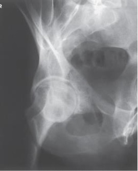
Definitions:
Occipital Lobe
The visual processing center of the mammalian brain, located in the back of the skull.
Cerebral Hemisphere
One of the two symmetrical halves of the brain, each of which has distinct functions but they work together to process complex cognitive tasks.
Visual Centers
Parts of the brain involved in processing visual information.
Corpus Callosum
A large band of neural fibers connecting the two brain hemispheres and carrying messages between them.
Q11: The central-ray angle for an AP axial
Q19: Letter B in the image below labels
Q21: The second largest tarsal bone, and the
Q24: For an axial projection of the calcaneus,
Q35: How far above the shoulders should
Q36: Which of the following joints should be
Q40: On which aspect of the foot does
Q51: Where is the central ray centered for
Q86: Where does the central ray enter
Q112: The PA oblique projection of the wrist