The figure below illustrates how a neuron with a myelinated axon transmits signals. 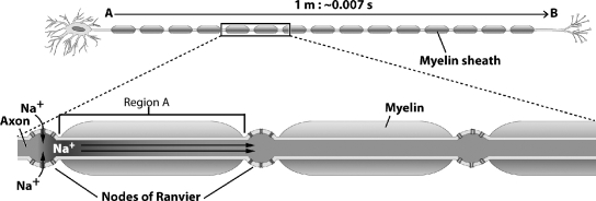
What is happening in Region A of this figure?
Definitions:
Perirhinal Region
A part of the brain involved in memory and recognition, located in the medial temporal lobe adjacent to the entorhinal cortex.
Grid Cells
Neurons located in the brain's entorhinal cortex that activate when an animal is at multiple locations forming a grid-like pattern, crucial for spatial navigation.
Place Cells
Hippocampal neurons maximally responsive to specific locations in the world.
Head Direction Cells
Neurons located in several brain regions that increase their firing rates when an animal's head points in a specific direction, aiding in spatial orientation and navigation.
Q6: Which of the following statements about gas
Q23: The figure below illustrates the inhalation step
Q25: A person who, as a result of
Q42: In the absence of _, nutrient recycling
Q43: When a plant germinates from a seed
Q44: Which of the following series correctly describes
Q48: After studying the image below, which of
Q54: Hormone replacement therapy (HRT)is used to ease
Q66: In which of the following animals would
Q76: Everyone who abuses drugs from an early