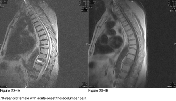
-In what imaging plane are Figure 20-4A and Figure 20-4B?
Definitions:
Venous Valves
Structures within veins that prevent the backward flow of blood, crucial for blood returning to the heart.
Tunica Media
The middle layer of the wall of a blood vessel consisting mainly of smooth muscle cells and elastic fibers, responsible for regulating the diameter of the vessel.
Lumen
The inside space of a tubular structure, such as a blood vessel or intestine.
Carotid Sinus
A dilated area at the base of the internal carotid artery that contains receptors to help regulate blood pressure.
Q1: Rupture of a bleb or bullae in
Q7: The most common cause for the radiographic
Q9: The typical sources of maduromycosis infection are
Q9: Which of the following is an occupational
Q12: If Paget disease was the underlying cause
Q13: What is the hallmark of giantism?<br>A) Generalized
Q13: Which statement best describes the function of
Q17: Which of the following diagnoses would be
Q25: Which diagnostic imaging modality is most appropriate
Q36: A 56-year-old male patient presents with new