West Nile virus (WNV) is a human pathogen that is transmitted by mosquitoes, which inject the virus into the host while they are feeding. To study the immune response to WNV, mice can be infected by injecting 100 PFU of virus into the footpad. This generates a robust immune response 7 days post-infection in the popliteal lymph node (LN), located behind the knee, as indicated in Figure. 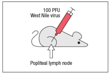
Popliteal LN cells are isolated from the mouse shown above at day 7 post-infection, and the T cells are stimulated in vitro with the WNV peptide P33 plus dendritic cells isolated from a non-infected mouse of the same strain. Five hours later, the CD8 T cells in the culture are analyzed for their ability to produce the cytokine IFN- , and the numbers of IFN- -producing CD8 T cells are quantified. As a control, T cells are also stimulated with an irrelevant non-viral peptide (ova) plus dendritic cells. The results are shown in Figure. 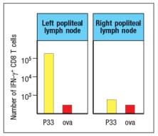
a) Why is the T cell response different between the two lymph node populations?
To identify the important antigen-presenting cell required to activate WNV-specific CD8 T cells in the popliteal LN, wild-type mice and Batf3-/- mice are each immunized with 100 PFU of WNV in the left footpad, as above. Previous studies have indicated that Batf3-/- mice lack one particular subset of conventional dendritic cells, known as CD8 + dendritic cells (DC), but otherwise appear to have normal numbers and subsets of all other immune cell populations (e.g., T cells, B cells, macrophages, etc.). The results of this experiment are shown in Figure
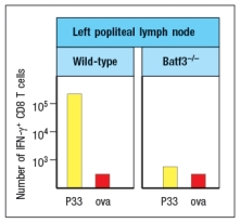
b) Name two possible functions of CD8 + dendritic cells that could account for the results seen in the Batf3-/- mice immunized with WNV.
To distinguish between the possible functions of CD8 + dendritic cells, another set of wild-type and Batf3-/- mice are immunized with 100 PFU of WNV in the left footpad. At day 7 post-infection, the left popliteal LN are isolated from the mice, and the CD4 T cells in these LN populations are tested for responses to a WNV peptide bound to MHC class II (peptide Y). As a negative control, an MHC class II-binding peptide from ova is used. These results are shown in Figure .
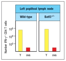
c) Do these results rule in/out either of your proposed functions of CD8 + dendritic cells indicated in answer to part (b)? Why or why not?
Definitions:
Soma
In the field of biology, it refers to the main structure of a living entity separate from its germ cells, and in neuroscience, it pertains to the neuron's soma.
Synapse
The small gap between two adjacent neurons, consisting of the presynaptic and postsynaptic neurons’ membranes and the space between them.
Dendrite
A projection of a neuron that receives signals (electrical and chemical) from other neurons or sensory cells, facilitating neural communication.
Axon
A long, slender projection of a neuron that conducts electrical impulses away from the neuron's cell body.
Q1: The skin and bodily secretions provide the
Q2: Antigen receptor signaling pathways are regulated by
Q3: Listeria monocytogenes is a bacterial pathogen that
Q4: Nitric oxide and superoxide radicals are toxic
Q6: Muckle-Wells syndrome is an autosomal dominantly inherited
Q8: T cells expressing <span class="ql-formula"
Q9: The pre-B-cell receptor provides an important
Q15: In some infectious diseases, antibodies specific for
Q29: When first discovered, investigators found it surprising
Q61: An index of clothing prices for 2014